The Brain’s Two-Origin Blueprint
 by ahsan
September 19, 2026
5 min read
by ahsan
September 19, 2026
5 min read
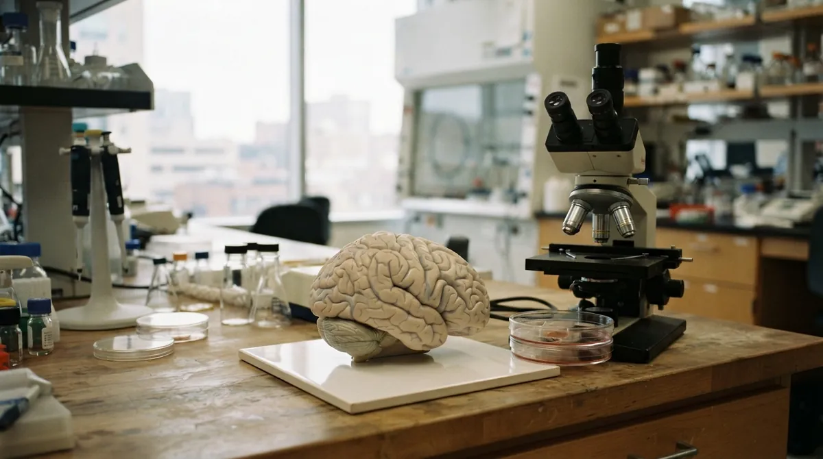
Picture an embryo before it has a recognizable head. During gastrulation, the early stage when cells rearrange into the body’s main layers, the future brain is only a sheet of tissue making decisions. A Stanford Medicine-led study published in Nature Neuroscience on September 18, 2026, suggests that one of those decisions is not to build a single brain from one master starting cell. Instead, two early cell populations begin separate journeys and later operate together inside one skull.
That is the careful translation of the striking headline that the human brain is two organs. The researchers are not describing two detached brains, two minds, or a new physical partition in adults. They are describing a developmental origin: one connected brain assembled from two lineage-restricted groups. A progenitor cell is an early cell that can produce a particular family of descendants, and lineage-restricted means its future options have already narrowed.
The headline needs a careful translation
The adult brain is usually divided into the forebrain, midbrain, and hindbrain. The forebrain contains much of the machinery for language, planning, memory, and conscious experience. The midbrain helps coordinate movement and sensory signals. The hindbrain includes important parts of the brainstem and cerebellum, supporting automatic functions such as breathing and heartbeat as well as motor coordination.
The study’s medical focus is more specific: hindbrain motor neurons, the nerve cells that help control muscles involved in facial movement, swallowing, and related functions. These cells matter in diseases such as spinal muscular atrophy (SMA), a genetic disease that damages motor neurons, and amyotrophic lateral sclerosis (ALS), a progressive disease that also destroys motor neurons. When these systems fail, swallowing and breathing can become dangerously difficult.
The split appears before the brain looks like a brain
To find the split, the researchers watched very early mouse embryos. They used lineage tracing, a method that labels cells and follows where their descendants end up. During gastrulation, two neural progenitor populations appeared in parallel:
 neural ectoderm
 / \
 anterior neural posterior neural
 ectoderm (Otx2) ectoderm (Gbx2)
 forebrain + midbrain hindbrain
Neural ectoderm is embryonic tissue that gives rise to the nervous system. The labels Otx2 and Gbx2 are genes whose activity acts as a molecular marker, meaning a measurable sign that helps researchers identify a cell population. The two groups did not overlap in the mouse embryos. One supplied the forebrain and midbrain; the other was committed to the hindbrain.
The team then tested the idea with human pluripotent stem cells. These are cells that can be guided into many different cell types in the body. When the researchers made anterior or posterior neural ectoderm-like cells, the two populations kept their separate tendencies: the anterior cells favored forebrain and midbrain identities, while the posterior cells favored hindbrain identities.
That mouse-and-human combination matters. The direct cell-tracking experiment was done in mice, while the human work used cells grown in the laboratory. Together, the results support a conserved developmental model without claiming that scientists have watched a living human embryo cell by cell.
Chromatin explains the dead end
The most revealing clue came from chromatin. Chromatin is the packaging made from DNA and proteins inside a cell. It controls which stretches of DNA are open for use and which are tucked away, much like a library where some books sit on the front desk while others remain in a locked archive.
Although nearly every cell carries the same DNA, its chromatin arrangement can make different genes available. The researchers found that anterior and posterior neural ectoderm had sharply different chromatin landscapes, meaning different patterns of accessible genetic material. Those patterns appeared early, before the cells had matured into recognizable neurons.
This helps explain a long-running problem in stem cell biology. Many lab protocols begin with neural progenitors that are already pointed toward a forebrain or midbrain fate, then try to steer them toward the hindbrain. The new results suggest that the obstacle was not a missing chemical signal at the end of the recipe. The cells had started on the wrong branch of the developmental tree.
Why have hindbrain neurons been so difficult to grow in the lab? The answer may begin several steps earlier than researchers expected.
A better recipe for hindbrain neurons
Once the team started from the posterior route, it was able to guide human pluripotent stem cells into hindbrain motor neurons associated with rhombomeres 5 and 6. Rhombomeres are temporary numbered segments that organize the developing hindbrain. The resulting cells were not merely shaped like neurons under a microscope. They produced action potentials, brief electrical signals that neurons use to communicate, and made proteins associated with hindbrain regions involved in facial and swallowing muscles.
That is an important technical distinction. A cell culture containing neurons is not the same thing as a living brain circuit. The cells lack the full arrangement of neighboring neurons, blood vessels, immune signals, and sensory inputs found in a person. Still, the researchers now have a human cell model that behaves like the specific hindbrain neurons they want to study.
For SMA and ALS research, that could change the experiments scientists can run. Researchers may be able to compare healthy and disease-associated neurons, observe how motor neurons become vulnerable, and test possible treatments in controlled cultures. Patient-derived stem cells could eventually make those comparisons more personal, although this study itself is a foundation for that work rather than a finished therapy.
An ancient blueprint
The two-origin pattern did not appear to be a peculiarity of mice or humans. The researchers found related evidence across animals including chickens, zebrafish, and acorn worms, small marine animals that are distant relatives of vertebrates. The authors propose that the arrangement may reach back roughly 550 million years, to a period when major animal lineages were taking shape.
Their evolutionary interpretation is intriguing: perhaps evolution did not design one giant nervous system from scratch. Instead, it may have brought two older neural programs together and gradually made them function as one connected system. That is an inference about deep history, not a claim that the human brain contains two independent control centers.
What this changes
The biggest shift is practical. Developmental biology often focuses on the final product—a neuron, a muscle cell, or an organ. This study argues that the starting population matters just as much. A cell cannot always be persuaded to become a type that its early chromatin state has ruled out.
For neuroscience, the result offers a better map of where the brain’s major regions come from and a more reliable route to making hindbrain neurons in the lab. It also gives researchers a sharper way to think about diseases that attack particular cell populations: vulnerability may depend partly on a cell’s earliest developmental identity.
The brain still functions as one connected organ. You do not carry two separate brains in your skull, and the study does not overturn the anatomy taught in medical textbooks. Its deeper message is more interesting: the brain’s unity is assembled. Long before thought, speech, or breathing circuits exist, two ancient developmental paths are already running side by side—and the cells remember which path they took.
Comments (0)
No comments yet. Be the first to respond!
Leave a Comment
Your comment will be visible after review.